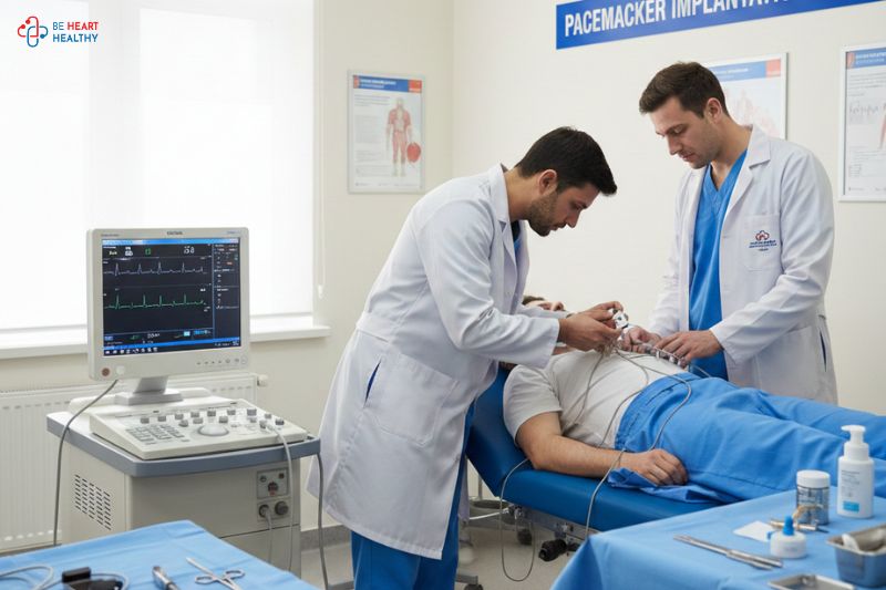
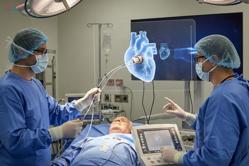
A non-invasive testing called 2D echocardiogram, sometimes known as 2D echo, is used to evaluate and examine the various parts of your heart. With the aid of sound waves, this test produces images of the various heart regions. It supports examining the damage, obstructions, and blood flow (1).
2-D (two-dimensional) echocardiography. Using this method, it is possible to “see” the heart’s true action. On the monitor, a cone-shaped 2-D echo image depicts the real-time motion of the heart’s structures. This enables the physician to observe and assess the various cardiac structures in action (1).
Clinical significance
It is advised for patients with cardiac issues to obtain a 2D Echo to check for any abnormalities in internal heart structures or heart function. Additionally, it can find birth problems in unborn children, such a hole in the heart. To diagnose and treat any heart problems early on and keep you fit and healthy as you age, doctors advise routine 2D echo exams (1).
Indications
To find the following heart problems, a 2D Echo is performed:
- any undiagnosed or untreated heart conditions
- inherited heart conditions, blood clots, and malignancies
- an issue with the heart valve
- abnormalities in the heart’s blood flow
- a slow blockage of the arteries caused by fatty compounds and other bloodstream contaminants.
- a cardiac enlargement brought on by thick or frail heart muscle
- Congenital heart disease. defects that appear in one or more cardiac components while the fetus is still developing, like a ventricular septal defect (the two lower chambers of the heart’s wall have a hole in them called the ventricular septal defect).
- Heart failure. a condition when blood cannot be pumped effectively because the heart muscle has grown weak or tight due to cardiac relaxation.
- An enlargement and weakening of the aorta or a portion of the heart muscle (the large artery that exits the heart and transports oxygenated blood to the body’s tissues).
- Heart valve disease. Dysfunction of one or more heart valves could result in irregular blood flow within the heart.
- Atrial or septal wall defects.Uneven pathways between the right and left sides of the heart might exist from birth, resulting from trauma, or develop during a heart attack.
- Shunts are a common finding in atrial and ventricular septal abnormalities, as well as when the liver and lungs drive erratic blood flow through the circulatory system.
Newer methods:
3-dimensional (3D) Echocardiography: Due to shortened exposures of the ventricles and atria in conventional 2D echocardiography, volumes can be underestimated (2). By allowing the user to choose images that are not foreshortened, 3D imaging removes the need for making geometric assumptions when calculating ventricular and atrial volumes. The visualization of the forms and spatial interactions between cardiac structures and the visualization and functionality of valves and valvular structures, can all be improved with 3D echocardiography (3).
All the latest information regarding heart issues and concerns is available online. One of the most popular websites that ensures to take best possible care for your heart is https://behearthealthy.in/contact-us/.
It has a wide range of useful topics providing new insights regarding cardiac care, heart health and cardiologist advice to live a happy, and joyful life. We care for you, so please reach out to us on our social media page for any query: https://www.facebook.com/careforyourheart.in/
References:
- https://www.hopkinsmedicine.org/health/treatment-tests-and-therapies/echocardiogram
- Tanabe K, Three-Dimensional Echocardiography - Role in Clinical Practice and Future Directions. Circulation journal: official journal of the Japanese Circulation Society. 2020
- Basman C,Parmar YJ,Kronzon I, Intracardiac Echocardiography for Structural Heart and Electrophysiological Interventions. Current cardiology reports. 2017


