Substantial improvements have been brought about in the techniques and tools used in the diagnosis and intervention of cardiovascular problems since the emergence of interventional cardiology. The complications have been relatively low and the success rate is remarkable in comparison to older tools and techniques. One such procedure used in interventional cardiology is Intravascular ultrasound (IVUS).
WHAT IS INTRAVASCULAR ULTRASOUND (IVUS)?
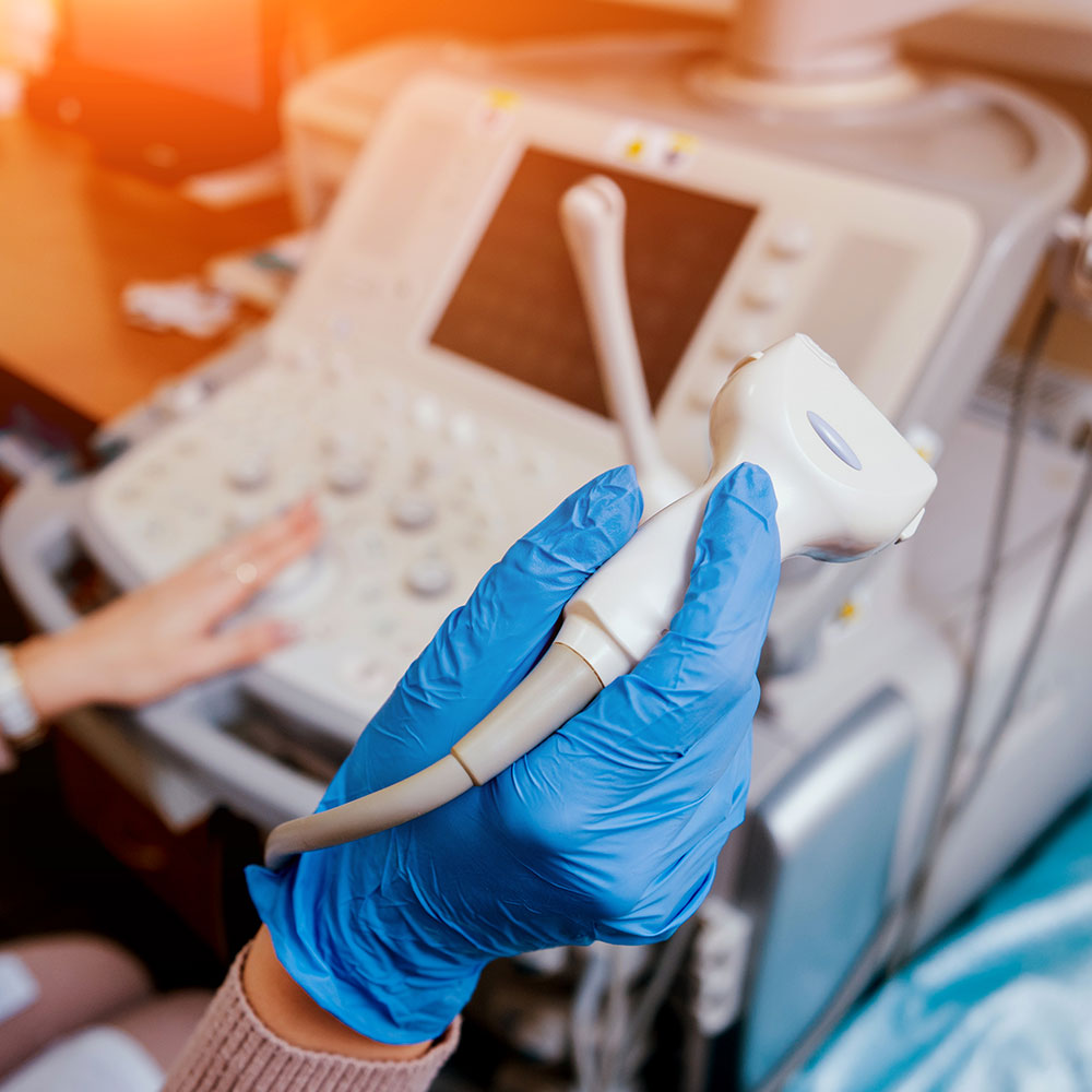
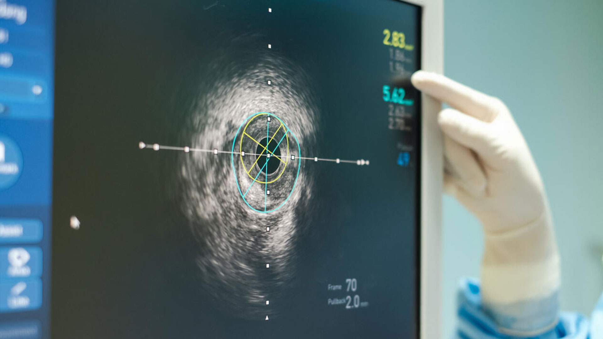
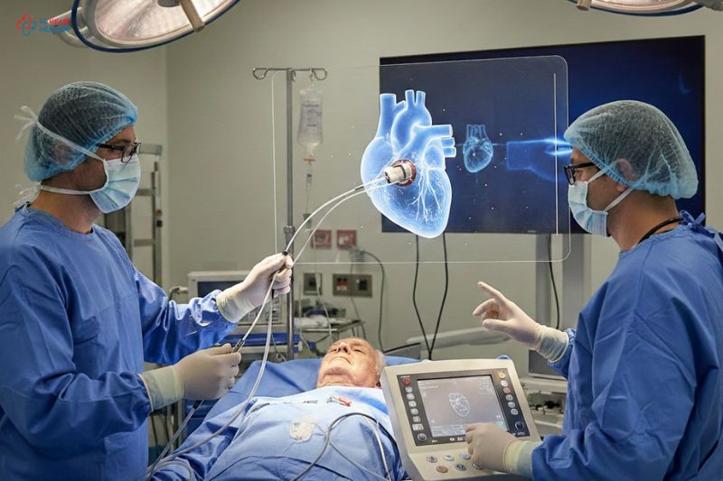
It is an imaging modality used primarily in interventional cardiology providing a pictorial view of what is going on inside the blood vessels. Sound waves are produced with the help of a probe or transducer that generates images of the blood vessels (1). A special catheter containing a small ultrasonic probe on one end is used in IVUS. This catheter is threaded through a vein or an artery by the doctor to the targeted place. Once it reaches the intended location, sound waves are emitted from the probe generating images of the blood vessels giving a 360-degree view of the vessels, and helping in assessment (2).
COMMON USES OF INTRAVASCULAR ULTRASOUND
Intravascular ultrasound assists doctors both in the diagnosis and treatment of various conditions of both veins and arteries. It helps doctors determine chronic and acute blood clots in veins especially when the cause is thought to be narrowing of veins. IVUS detects the blocked or narrowed vessels deep in the body. Furthermore, IVUS aids in the measurement of veins for the selection of appropriate stent size (3).
Intravascular ultrasound has also proved to be useful in the assessment of the body’s arteries. It helps doctors visualize coronary arteries and peripheral leg arteries. IVUS is often used by doctors in combination with catheter angiography in the diagnosis of peripheral artery disease (PAD). Also, the size of the stent to be used in PAD to keep the arteries open is determined with the help of IVUS (4).
The visualization of coronary arteries can be carried out with IVUS in conjunction with angioplasty, angiography, and stent placing or it aids in the planning of these procedures. The entire wall of arteries becomes visible with IVUS, unlike angiography. This helps in disclosing more information about the buildup of plaque (atherosclerosis). The chances of heart attack increase due to plaque formation. The information obtained through IVUS affects the decisions in the treatment strategy. For example, the place and size of the stent. Often after vascular stenting and angioplasty, IVUS is used to confirm the correct placing of the stent and that the problem has been resolved (5). Moreover, abdominal aortic aneurysm is also assessed by IVUS before the intervention and during and after the repair of the vessel, helps is taken from IVUS.
THE HEART SPECIALIST IN MUMBAI – CARE FOR YOUR HEART
Care for Your Heart is equipped with the latest equipment and technologies. Dr. Ankur Phatarpekar and his team of expert professionals at Care for Your Heart specialize in interventional cardiology and coupled with their expertise and experience, you can get the best treatment plan for your heart. For further information and guidance contact CARE for YOUR HEART.

Source
1. Parviz Y, Shlofmitz E, Fall KN, Konigstein M, Maehara A, Jeremias A, et al. Utility of intracoronary imaging in the cardiac catheterization laboratory: comprehensive evaluation with intravascular ultrasound and optical coherence tomography. Br Med Bull. 2018 Mar 1;125(1):79–90.
2. Shlofmitz E, Kerndt CC, Parekh A, Khalid N. Intravascular Ultrasound. In: StatPearls [Internet]. Treasure Island (FL): StatPearls Publishing; 2022 [cited 2022 Mar 8]. Available from: http://www.ncbi.nlm.nih.gov/books/NBK537019/
3. Weissman NJ, Mintz GS. Chapter 7 – Intravascular Ultrasound: Principles and Clinical Applications**See page 169 for a list of key acronyms for this chapter. In: Otto CM, editor. The Practice of Clinical Echocardiography (Third Edition) [Internet]. Philadelphia: W.B. Saunders; 2007 [cited 2022 Mar 8]. p. 152–71. Available from: https://www.sciencedirect.com/science/article/pii/B9781416036401500113
4. Gagne PJ, Bowman J, Kucher T. Chapter 16 – The Role of IVUS in Arterial and Venous Procedures in the Office-Based Laboratory. In: Jain KM, editor. Office-Based Endovascular Centers [Internet]. Elsevier; 2020 [cited 2022 Mar 8]. p. 119–26. Available from: https://www.sciencedirect.com/science/article/pii/B9780323679695000162
5. Drakopoulou M, Toutouzas K, Tousoulis D. Chapter 2.5 – Acute Coronary Syndromes. In: Tousoulis D, editor. Coronary Artery Disease [Internet]. Academic Press; 2018 [cited 2022 Mar 8]. p. 201–33. Available from: https://www.sciencedirect.com/science/article/pii/B978012811908200012X


