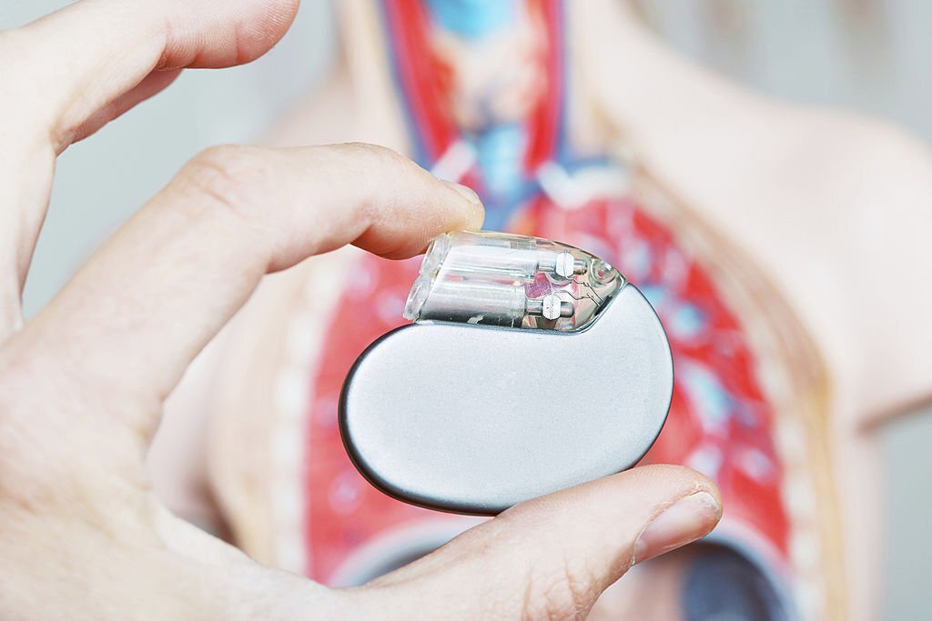
What is a pacemaker?
The sinus node, which acts as the heart’s pacemaker, is where the normal cardiac activity starts. After crossing the atria to reach the atrioventricular (AV) node, electrical wavefronts quickly spread to and depolarized the ventricles by entering the His-Purkinje system. When intrinsic cardiac automaticity or conduction integrity fails, the electrical excitability of cardiac tissue enables a small, external electrical stimulus to drive myocytes to the threshold, resulting in the depolarization of nearby myocytes through biological processes that consume energy and the subsequent propagation of an electrical wave front, with nearly simultaneous muscular contraction via excitation-contraction coupling. Pacemakers deliver this outside stimulus1. In another word, artificial pacemakers are electronic devices that use electrical impulses to stimulate the heart to keep or restore a regular rhythm in those with bradyarrhythmia.
How do they function?
Flexible insulated cables, or leads, are used to transmit electrical impulses from the pacemaker’s generator to the heart muscle and to return data about the heart’s normal operations. The right atrium and right ventricle are the most frequent locations for these wires, or leads, to be implanted within the heart. One kind of pacemaker is “wireless”.
Most often, the pacing lead contains one or two electrical “poles.” When necessary, an electrical impulse is sent to the heart muscle, and the lead can detect the electrical activity of the heart itself.
For whom is it done?
A pacemaker is most frequently used for bradyarrhythmia. The choice of whether to employ one of these devices and which kind, in particular, will rely on many variables, including:
- Whether the condition is short-term or long-term
- The existence or absence of the symptoms related to arrhythmia (syncope, presyncope, palpitations etc.)
- The precise characteristics and underlying reasons for the arrhythmia
- The predicted pacer’s frequency
- the fundamental cardiac issues
Types of pacemakers
Different pacemaker designs and pacing techniques have been created to restore or maintain a normal heartbeat in various ways. Additionally, pacemakers can have one, two, or three chambers:
- One lead is used by single-chamber pacemakers to send and receive impulses to and from the right atrium or right ventricle.
- The two leads that a dual-chamber pacemaker typically has, one to the right atrium and one to the right ventricle, can enable a cardiac rhythm that more closely resembles the regular activity of the heart and reflects intrinsic depolarization.
- A right atrium lead, a right ventricle lead, and a left ventricle lead are commonly present in triple-chambered pacemakers. Patients who have compromised cardiac musculature use these devices (which results in heart failure). These pacemakers “resynchronize” the ventricles, which might increase the effectiveness of the heart’s contraction. They are also known as “biventricular pacing”
Temporary pacemakers are designed for transitory use while in the hospital. They are employed either because it is anticipated that the arrhythmia would pass temporarily and eventually, or because the patient needs short-term care until a permanent pacemaker can be implanted. Pacemakers that are designed for long-term usage are called permanent pacemakers.
How is it done?
Pre-pectoral implantation refers to the placement of the pacemaker in the soft tissue beneath the skin in the region below the clavicle, which is above the pectoral muscle but beneath the skin and adipose tissue. Typically, the pacemaker leads are placed transvenous into a large vein and advanced until they are firmly implanted within the necessary area(s) of the heart muscle. The pulse generator is connected to the other ends of the leads. The pulse generator may occasionally be positioned beneath the skin of the upper abdomen.
In most cases, a specialist (cardiologist, surgeon, or cardiac electrophysiologist) with experience in this process implants the pacemaker in a sterile laboratory or operating room. The surgery is as painless as possible thanks to the use of local anesthesia and frequently conscious sedation. Only rarely general anesthesia is necessary. Utilizing X-ray imaging, the location of the pacemaker leads is often examined (called fluoroscopy). The kind of device that is being implanted will determine how long the surgery takes. Without a separate pulse generator, leadless pacemakers are often implanted through a leg vein and put in the heart muscle. This can be done as an outpatient procedure, and hospital stays are often minimal. Although there may be some limitations on arm movement and activities for the first two to four weeks after the treatment, recovery is swift. Pacemakers can be configured to alter the resting heart rate, the maximum heart rate at which they will pace, and the changes in heart rate that should happen during physical activity once they have been implanted.
Conclusion
From the development of totally implantable devices to the leadless pacemakers, artificial cardiac pacemakers have embarked on a unique journey that has not reached their destination yet as their advances of it continue to date.
References
- Mulpuru SK, Madhavan M, McLeod CJ, Cha YM, Friedman PA. Cardiac Pacemakers: Function, Troubleshooting, and Management: Part 1 of a 2-Part Series. J Am Coll Cardiol. 2017;69(2):189-210. doi:10.1016/J.JACC.2016.10.061
- Patient education: Pacemakers (Beyond the Basics) – UpToDate. Accessed August 25, 2022. https://www.uptodate.com/contents/pacemakers-beyond-the-basics/print


